Anterior Cruciate Ligament Injury
by Tahir Mahmud
Introduction
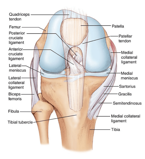
The anterior cruciate ligament (ACL) is a ligament that provides stability to the knee joint. It is commonly injured during high-intensity sports. The ACL is commonly torn by either a hyper-extension injury or a pivoting injury to the knee joint. Surgery is often recommended to restore knee stability and function by reconstructing a damaged ACL with a graft. The graft may be obtained from various locations around the patient’s own knee (Autograft) including the hamstring tendons, the tendon that stabilizes the kneecap or patella called a “bone-patellar tendon-bone” graft, the quadriceps tendon, or from donated tissue (Allograft).
Disease Overview
The thigh bone and shin bone are connected by a group of ligaments that provide stability to the knee. These include the anterior and posterior cruciate ligaments that run through the centre of the knee and cross each other diagonally. Together, they function to control the back and forth movements of the knee. The ACL, situated more in front, prevents the shin from slipping forward and provides stability during rotational movements. Intense stress on the ACL due to accidents and sports injuries may cause it to stretch or tear. ACL injuries may also be accompanied by injury to articular cartilage (joint surface), meniscal cartilage (shock absorber), or other ligaments and tissues in the joint.
Indications
Knee injuries that involve the ACL usually cause complete or near complete tears that require surgical reconstruction with a graft to prevent instability and restore function to the knee. ACL reconstructive surgery is recommended for those who wish to return to a high level of multi-directional activity.
The diagnosis in confirmed with careful clinical examination and the aid of a MRI scan.Injuries in addition to the ACL may be confirmed on the MRI scan.
Surgical procedure
ACL reconstruction is usually performed 3-8 weeks after an injury once swelling has reduced and range of movement is improved through rehabilitation. This prevents stiffness and scar formation after surgery.
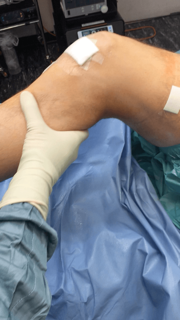
The procedure is performed under general or local anaesthesia. Your doctor will first evaluate your knee under anaesthesia to assess the damage to the ACL and other structures.
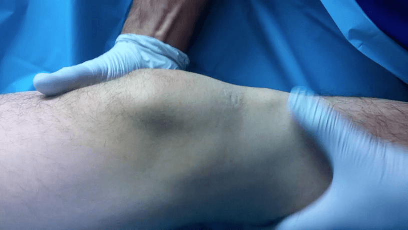
Following this, the new ACL graft tissue is harvested. This picture shows the hamstring tendons harvested for a ACL reconstruction using a minimally invasive technique.
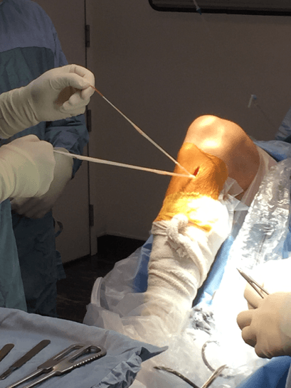
Small incisions (surgical cuts) are then made over the knee to insert an arthroscope and other surgical instruments. The arthroscope consists of a tube with a light and camera to view the inside of the joint. The structures within the knee are evaluated and any significant injuries are identified and repaired during the surgery. Tunnels are created by drilling through the shin bone and thigh bone. The graft is passed through these tunnels and secured with screws to replace the original ACL. The incisions are then closed with sutures and covered with dressings. The procedure takes about 1 hour.
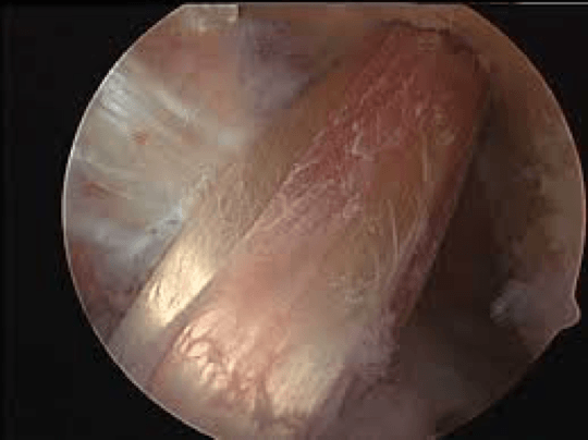
Arthroscopic image of new ACL graft in place.
Post-Operative Care
Following surgery, you will have some pain and swelling for which your doctor will prescribe medications to keep you comfortable. Ice application and leg elevation is recommended to control swelling. Knee movement and weight bearing are encouraged as soon as possible. Physical therapy will be recommended to improve strength and movement.
Summary
Most ACL injuries require reconstruction with a suitable tendon graft to restore function and stability.
